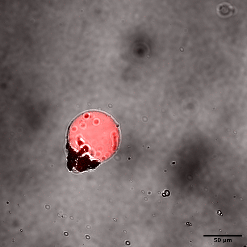
The image is of liposomes that we were able to actually create. We conducted fluorescence observation using confocal microscopy. The observation was carried out with a laser at an excitation wavelength of 559nm. The red fluorescence of rhodamine contained in the IS reveals that the liposomes exist as spherical membrane structures. As can be seen from the image, there are parts where the membrane surface of the liposome is not in a clean state. We believe this is due to the effects of the interface passage method. As a countermeasure, we could consider increasing the number of times the liposomes are washed.